
1. 서론
This manual provides essential instructions for the safe and effective use of your Ken-A-Vision T-17541C Digital CoreScope 2 Compound Microscope. Designed for biological and educational applications, this digital microscope features a monocular head, LED illumination, and a 1.3-megapixel camera for capturing still images and video. Please read this manual thoroughly before operating the device and retain it for future reference.
2. 안전 정보
- 전기 안전: Ensure the power supply is connected to a grounded outlet. Do not operate the microscope with wet hands or in damp 환경. 청소나 정비를 하기 전에 전원을 분리하세요.
- 손질: Always carry the microscope by its base and arm. Avoid sudden impacts or vibrations.
- 안과 진료: Do not touch optical surfaces with bare fingers. Use only approved lens cleaning materials.
- 통풍: 현미경의 과열을 방지하기 위해 적절한 환기가 이루어지도록 하십시오.
- 어린이들: This device is not a toy. Adult supervision is recommended when used by children.
3. 패키지 내용
아래 나열된 모든 품목이 패키지에 들어 있는지 확인하세요.
- Ken-A-Vision CoreScope 2 Digital Microscope
- 전원 공급 장치
- Applied Vision 4 Software (for Windows, Mac, and Linux)
- Microscope Cover
- 앨런 렌치
- 사용 설명서(이 설명서)
4. 제품 오버view
The Ken-A-Vision T-17541C CoreScope 2 is a robust digital compound microscope designed for clarity and ease of use. Key components include:
- Monocular Head: Features a fixed 45-degree incline for comfortable viewing and 360-degree rotation.
- 접안 렌즈: 10x widefield eyepiece with a pointer for easy observation.
- 목표: Reverse-mounted 4x, 10x, and 40xS DIN achromatic lens objectives, protected against humidity and climate variations.
- Floating Stage: 라운드 stage with spring-loaded clips to secure slides and a stop mechanism to prevent damage.
- 초점 조절 노브: Separate coaxial coarse and fine focus controls for precise adjustments.
- LED 조명: Built-in bright and cool white LED light with dimmer and on/off switch.
- 디지털 카메라: Integrated 1.3-megapixel (MP), 720P high-definition (HD) CMOS camera with a built-in USB port for direct connection to a computer or monitor.

그림 1: 정면 view of the Ken-A-Vision T-17541C Digital CoreScope 2 Compound Microscope, showing the monocular head, objective lenses, floating stage, focus knobs, and connected USB cable.
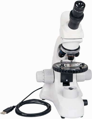
그림 2: 각진 view of the Ken-A-Vision T-17541C Digital CoreScope 2 Compound Microscope, highlighting the compact design and the position of the objective lenses and stage.
5. 설정
- 짐 풀기: 현미경과 모든 부속품을 포장에서 조심스럽게 꺼내십시오.
- 놓기: 현미경을 직사광선, 과도한 열 또는 진동을 피해 안정적이고 평평한 곳에 놓으십시오.
- 전원 연결: Connect the provided power supply to the microscope and then plug it into a standard electrical outlet (120-240V). The microscope can also be powered via a low voltage USB computer connection.
- 소프트웨어 설치: For digital imaging, install the Applied Vision 4 software on your computer. The software is compatible with Windows (XP SP2 or later, Vista, 7), Mac (OS 10.5 or later), and Linux. Follow the on-screen instructions during installation.
- USB 연결: Connect the microscope's built-in USB port to your computer using a USB cable. This connection enables the digital camera functionality and can also provide power.
- 커버 제거: Remove the microscope cover before use.
6. 사용 설명서
- 전원 켜기: Turn on the microscope using the on/off switch for the LED illumination.
- 조명 조정: Use the dimmer control to adjust the brightness of the LED light to a comfortable level for viewing your specimen.
- 표본 채취 장소: Place your prepared slide on the floating stage, securing it with the spring-loaded clips.
- Select Objective: Rotate the nosepiece to select the desired objective lens (4x, 10x, or 40xS). Start with the lowest magnification (4x) for initial view주고받음.
- 초점:
- 사용하세요 coarse focus knob (larger knob) to bring the specimen into approximate focus.
- 그런 다음 다음을 사용합니다. fine focus knob (smaller knob) for precise focusing and to achieve a sharp image.
- 접안렌즈 조정: Adjust the variable diopter on the eyepiece for optimal visual clarity.
- Rotate Head: The monocular head can be rotated 360 degrees for shared viewing or comfortable positioning.
- Digital Imaging (with software):
- Ensure the microscope is connected to your computer via USB and the Applied Vision 4 software is running.
- The software will display the live feed from the 1.3 MP HD CMOS camera.
- Use the software interface to capture still images or record video. Refer to the Applied Vision 4 software manual for detailed instructions on its features.
- 전원 끄기 : When finished, turn off the LED illumination and disconnect the power supply. Cover the microscope with the provided dust cover.
7. 유지관리
- 본체 청소: 현미경 본체를 부드러운 천으로 닦으십시오.amp 옷감. 가혹한 화학 물질이나 용제를 사용하지 마십시오.
- 렌즈 세척: Use a soft lens brush to remove dust. For smudges, use a specialized lens cleaning solution and lens paper. Do not use abrasive materials.
- 저장: Always cover the microscope with the dust cover when not in use to protect it from dust and debris. Store in a dry environment.
- 습도 조절: The objective lenses are designed with humidity and climate control protection, but prolonged exposure to extreme conditions should be avoided.
8. 문제 해결
- 조명 없음: Check if the power supply is properly connected and the on/off switch is engaged. Ensure the dimmer is not set to its lowest setting.
- 이미지가 흐릿합니다. Adjust both the coarse and fine focus knobs. Ensure the objective lens is properly clicked into place. Clean the eyepiece and objective lenses if smudges are present.
- No Image on Computer Screen: Verify the USB cable is securely connected to both the microscope and the computer. Ensure the Applied Vision 4 software is installed and running correctly. Check your computer's device manager to confirm the camera is recognized.
- Specimen Not Centered: Gently adjust the position of the slide on the floating stage.
9. 사양
| 특징 | 사양 |
|---|---|
| 모델 번호 | 티-17541C |
| View구성 | Monocular with 45-degree incline, 360-degree rotation |
| 접안 렌즈 | 10x widefield with pointer |
| 대물렌즈 | 4x, 10x, 40xS DIN Achromatic |
| 최대 배율 | 400배 |
| 조명 | Bright and cool LED white light with dimmer and on/off switch |
| 집중하다 | 동축식 조대 및 미세 초점 조절 |
| 디지털 카메라 | 1.3 MP, 720P HD CMOS, USB 2.0 |
| 소프트웨어 호환성 | Applied Vision 4 (Windows XP SP2+, Vista, 7; Mac OS 10.5+; Linux) |
| Stage | Round floating stage with 2 spring-loaded clips |
| 전원 | 낮은 볼륨tage USB computer connection or external USB power plug, 120-240V |
| 재료 | 알류미늄 |
| 제품 크기(높이 x 너비 x 깊이) | 40.0 x 30.48 x 22.86cm(15.75 x 12 x 9인치) |
| 무게 | 2.31kg(5.1파운드) |
| 인증서 | ISO:9001 – 2000, CE, CSA, RoHS |
10. 보증 및 지원
The Ken-A-Vision T-17541C Digital CoreScope 2 Compound Microscope is manufactured by Ken-A-Vision Manufacturing, headquartered in Kansas City, MO. For warranty information, technical support, or service inquiries, please refer to the official Ken-A-Vision website or contact their customer service department directly. Please have your model number (T-17541C) and purchase date available when contacting support.